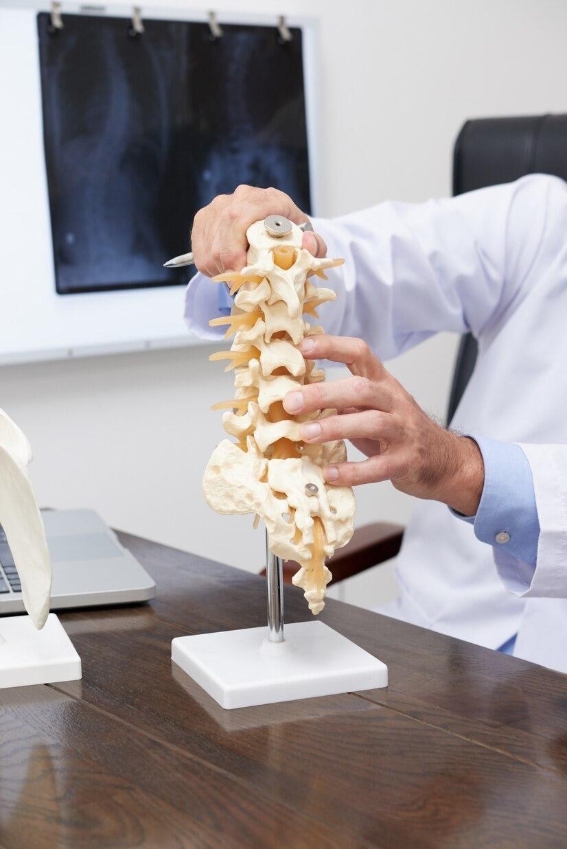
Spinal disorders, ranging from degenerative conditions to acute injuries, can significantly impact quality of life, often leading to chronic pain, mobility restrictions, and neurological deficits. For many years, surgical intervention for these conditions typically involved extensive “open” procedures, characterized by large incisions, significant muscle dissection, and prolonged recovery periods. However, the advent of minimally invasive spine surgery (MIS) has revolutionized the field, offering patients effective treatment options with significantly reduced collateral damage and faster rehabilitation.
MIS represents a paradigm shift in surgical philosophy. Instead of large incisions that cut through muscles, MIS techniques utilize small incisions, specialized instruments, and advanced imaging guidance to access the spine. The core principle is to achieve the surgical objective – whether decompression of nerves, stabilization of a segment, or repair of a fracture – while minimizing trauma to surrounding healthy tissues. This approach translates into numerous benefits for patients, including less blood loss, reduced postoperative pain, shorter hospital stays, and a quicker return to daily activities.
The Core Principles of Minimally Invasive Spine Surgery
At the heart of MIS lies the commitment to precision and tissue preservation. Surgeons employ a variety of specialized tools and techniques:
- Small Incisions: Rather than a single long incision, MIS uses one or a few small incisions, typically 1 to 2 centimeters in length.
- Tubular Retractors: These narrow tubes are gently inserted through the small incision, displacing muscle tissue rather than cutting it. Dilators are used sequentially to gradually expand the opening to the desired diameter, creating a working channel to the spine.
- Endoscopes and Microscopes: High-definition endoscopes provide magnified, illuminated views of the surgical field on a monitor, while operating microscopes offer stereoscopic, magnified visualization for intricate work.
- Specialized Instruments: Long, thin instruments are designed to fit through the tubular retractor or small incisions, allowing the surgeon to perform delicate maneuvers with precision.
- Intraoperative Imaging: Fluoroscopy (real-time X-ray), O-arm navigation, and CT scans provide surgeons with precise, real-time guidance, ensuring accurate placement of implants and precise tissue removal.
While the fundamental goals of surgery remain the same, the application of these principles allows for a highly targeted approach. Let us delve deeper into how MIS techniques are applied to specific spinal conditions.
A Deeper Dive into Specific Conditions
1. Herniated Discs: Lumbar Microdiscectomy
A herniated disc occurs when the soft, jelly-like center of an intervertebral disc pushes through a tear in its tougher outer layer, often compressing nearby spinal nerves. This can lead to severe pain, numbness, or weakness in the back and legs (sciatica).
MIS Approach (Lumbar Microdiscectomy): This is one of the most common and successful MIS procedures. Through a small incision (typically less than an inch) in the lower back, a tubular retractor is guided to the affected disc. Using an operating microscope or endoscope for magnified visualization, the surgeon carefully removes only the herniated portion of the disc that is impinging on the nerve. The majority of the healthy disc is left intact.
Benefits: The primary advantages are minimal muscle disruption, less blood loss, and significantly reduced postoperative pain compared to open discectomy. Patients often go home the same day or the following morning and can resume light activities much faster. The success rates for pain relief are comparable to open surgery, but with a more favorable recovery profile.
2. Spinal Stenosis: Lumbar Decompression (Laminectomy/Foraminotomy)
Spinal stenosis refers to the narrowing of the spinal canal or the nerve root passages (foramina), which can compress the spinal cord or nerves. This narrowing is most commonly caused by age-related degeneration, bone spurs, thickened ligaments, or bulging discs, leading to symptoms like leg pain, numbness, and weakness, especially with walking (neurogenic claudication).
MIS Approach (Minimally Invasive Decompression): Instead of removing large sections of bone (lamina) and muscle to access the entire spinal canal, MIS decompression focuses on targeted removal. Using tubular retractors and microscopic or endoscopic visualization, the surgeon precisely removes only the bone, ligament, or disc material that is causing the compression. This might involve a unilateral laminotomy (removing a small portion of the lamina on one side) or a foraminotomy (enlarging the nerve root opening).
Benefits: By preserving more of the posterior spinal structures (bone and ligaments), MIS decompression helps maintain spinal stability, reducing the need for concurrent fusion in some cases. It results in less postoperative pain, a lower risk of infection, and a quicker return to mobility compared to open laminectomy.
3. Spinal Instability and Spondylolisthesis: Minimally Invasive Spinal Fusion
Spinal fusion is a surgical procedure designed to permanently connect two or more vertebrae, eliminating motion between them. It is typically performed to alleviate pain caused by spinal instability, such as severe degenerative disc disease, spondylolisthesis (where one vertebra slips forward over another), or conditions that cause deformity.
MIS Approach (MIS TLIF/PLIF – Transforaminal/Posterior Lumbar Interbody Fusion): Performing spinal fusion minimally invasively is significantly more complex than decompression or discectomy but offers substantial advantages. In a MIS TLIF or PLIF, multiple small incisions are made in the back. Percutaneous (through the skin) screws are precisely guided into the vertebrae using real-time imaging (fluoroscopy or navigation). A tubular retractor is then used to access the disc space, allowing the surgeon to remove the damaged disc and insert a bone graft or a cage filled with bone graft material. Rods are then connected to the percutaneous screws to stabilize the segment while the fusion occurs.
Benefits: The main advantage is the avoidance of extensive muscle stripping, which is a major source of pain and morbidity in traditional open fusions. This translates to significantly less blood loss, reduced postoperative pain, shorter hospital stays, and a faster recovery. While the fusion rate is comparable to open fusion, patients often experience an easier and less painful rehabilitation process.
4. Vertebral Compression Fractures: Vertebroplasty and Kyphoplasty
Vertebral compression fractures (VCFs) are common, particularly in elderly patients with osteoporosis, and can cause severe, debilitating back pain. They occur when a vertebra collapses, often due to weakened bone, or sometimes due to trauma or tumors.
MIS Approach (Vertebroplasty/Kyphoplasty): These are inherently minimally invasive, percutaneous procedures.
- Vertebroplasty: Under imaging guidance, a small needle is inserted through a tiny skin incision directly into the fractured vertebra. Bone cement (polymethyl methacrylate, PMMA) is then injected into the compressed bone to stabilize it and alleviate pain.
- Kyphoplasty: Similar to vertebroplasty, but a balloon is first inserted through the needle and inflated within the fractured vertebra to create a cavity and, often, restore some of the vertebral body height before the bone cement is injected.
Benefits: Both procedures are performed through very small punctures, often under local anesthesia and sedation. They offer rapid pain relief, often within hours or days, by stabilizing the fracture. Patients can typically return home the same day and experience a dramatic improvement in their quality of life.
Overarching Advantages of MIS
Beyond the condition-specific benefits, MIS procedures collectively offer several overarching advantages:
- Reduced Muscle Damage: The key differentiator, leading to less pain and faster functional recovery.
- Minimal Blood Loss: Smaller incisions and less tissue disruption result in significantly less intraoperative bleeding, reducing the need for blood transfusions.
- Lower Risk of Infection: Smaller wounds inherently carry a lower risk of surgical site infections.
- Reduced Postoperative Pain: Less tissue trauma generally means less pain, often requiring fewer strong pain medications.
- Shorter Hospital Stays: Many MIS procedures are outpatient or require only a one-night stay.
- Faster Rehabilitation: Patients can typically start physical therapy sooner and return to their normal activities more quickly.
- Reduced Scarring: Smaller incisions leave less noticeable scars.
Patient Suitability and Considerations
While the benefits of MIS are compelling, it is crucial to understand that not all spinal conditions or all patients are candidates for minimally invasive approaches. Complex deformities, multi-level severe instability, extensive tumors, or severe infections might still necessitate traditional open surgery.
Patient selection is paramount. A comprehensive evaluation by an experienced spine surgeon is essential to determine the most appropriate surgical approach. Factors considered include the specific diagnosis, the severity of the condition, unique patient anatomy, overall health, and prior surgical history. The expertise of the surgical team, including specialized training in MIS techniques and access to advanced equipment, is also critical for optimal outcomes.
The Evolving Landscape of MIS
The field of minimally invasive spine surgery continues to evolve rapidly. Advancements in imaging technology, such as intraoperative navigation systems and robotics, are making MIS procedures even more precise and safer. Augmented reality and virtual reality technologies are also beginning to play a role in surgical planning and execution. New developments in instrumentation, bone graft materials, and surgical techniques are constantly expanding the scope of conditions that can be treated minimally invasively.
Conclusion
Minimally invasive spine surgery has transformed the treatment landscape for a wide array of spinal conditions, offering patients a less invasive, less painful, and more efficient path to recovery. By leveraging small incisions, specialized instruments, and advanced imaging, surgeons can effectively address conditions like herniated discs, spinal stenosis, instability, and vertebral fractures with significantly reduced impact on surrounding tissues. While not a panacea for all spinal ailments, MIS represents a powerful and continually advancing frontier in modern spine care, allowing many individuals to regain function and a better quality of life with remarkable speed and comfort.


